Strong steels with outstanding mechanical properties are integral for various technological applications. However, the formation of those steels has been traditionally limited by alloying with additives, involving stringent requirements of rapid quenching and complex aging.
Despite tremendous efforts over decades, the strengthening of ferrous steels with low-concentration additives is still challenging, especially for pure iron (Fe). This difficulty stems from the lack of forming spatial obstacles, such as martensites and precipitates, which are indispensable ingredients for mechanical enhancement. In addition, in the absence of a clean system for studying the martensitic transition, the underlying transition mechanism—a longstanding issue that has eluded physicists for centuries—is still unclear.
A research team led by Associate Professor Shanmin Wang from the Department of Physics at the Southern University of Science and Technology (SUSTech) and the Quantum Science Center of Guangdong-Hong Kong-Macao Greater Bay Area has recently achieved progress on the study of mechanical enhancement of pure Fe by the formation of hierarchically structured martensites using high-pressure (P) techniques that are combined with nanoengineering.
Their experiments have led to the discovery of ultrastrong pure iron with excellent mechanical properties exceeding those of high-speed steels (HSS). Based on the simplest system of pure Fe, they have gained valuable insights into the martensitic transition mechanism.
Their work, entitled “Formation of hierarchically structured martensites in pure iron with exceptional strength and stiffness”, was published in the high-impact academic journal Proceedings of the National Academy of Sciences of the United States of America (PNAS).

Figure 1. (A) Lattice of a pure metal and dislocation slip along certain crystallographic planes under stress. (B) Different types of obstacles (Science 324, (2009) 349).
Pure iron, the primary component of ferrous steels, is mechanically soft and highly ductile due to the ease of dislocation slips along certain slip systems under stress (Figure 1A). To obtain strong steels, high concentrations of carbon or other alloying elements are often required to drive phase transitions, such as martensitic transition. These transitions produce high-density spatial obstacles, including interstitial atoms, precipitates, dislocations, grain boundaries, and twin boundaries (Fig. 1B). However, achieving such phase transitions without the involvement of solute atoms is exceedingly difficult in pure Fe, which impedes the development of high-strength, low-alloy steels.
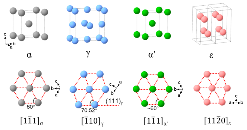
Figure 2. Polytypes of Fe. The phases of α, γ, α´, and ε are the ground-state, high-temperature, martensitic, and high-pressure structures, respectively.
The mechanical properties of steel are closely linked by Fe’s ability to exist in various crystal structures: body-centered cubic (bcc) α, face-centered cubic (fcc) γ, body-centered tetragonal (bct) α´, and close-packed hexagonal (hcp) ε phases (Figure 2). Upon cooling or straining, stacking faults (SFs) are nucleated and induce structural transitions, such as γ-Fe transforming to other polytypes through relative shifts of atomic planes. Alloying with solute atoms alters these transition paths, creating features like nanotwins and precipitates that act as obstacles to dislocation movement. These obstacles often form multiscale martensites, further strengthening the material. However, the complexity of martensitic transitions (i.e., high-temperature γ phase to martensitic α´ phase) in conventional steels—relying on various additives and stringent treatment processes—leaves the associated transition mechanism unclear, despite different proposed models.
Recent experiments showed that high-pressure conditions favor martensitic transition in steels without the requirement of rapid quenching, as stacking fault energy decreases under high-pressure and high-temperature conditions. Spatial confinement also provides an alternative approach for kinetically stabilizing metastable Fe polytypes using nano-effect, altering transition pathways. Additionally, nanostructured materials with dense grain boundaries provide active sites for partial dislocation nucleation under stress. These dislocations promote stacking fault formation and drive multiple-phase transitions, ultimately creating a hierarchy of nanoscale obstacles that significantly strengthen the material.

Figure 3. (A) Cubic module of large-volume press (DS 6×10 NM) to generate high-pressure and high-temperature conditions for preparing ultrastrong m-Fe samples. (B) SEM image of starting nanocrystalline Fe. Each particle is an aggregation of nanoparticles of Fe with a characteristic size of ~87 nm. (C) Experimental setup of in situ high-pressure and high-temperature energy-dispersive synchrotron XRD measurements at the beamline of 6BM-B/APS.
Using advanced high-pressure techniques and nanocrystalline Fe powders as starting materials (Figure 3A-3B), the research team created ultrastrong pure-Fe bulk samples with superior hardness, strength, and elasticity compared to high-speed steels (HSS). Postmortem analysis of these samples revealed the mechanisms behind their enhanced mechanical properties. Additionally, in situ high-pressure and high-temperature synchrotron XRD measurements (Figure 3C) captured the structural evolution of Fe under varying high-pressure and high-temperature conditions, shedding light into the kinetics of martensitic transitions.
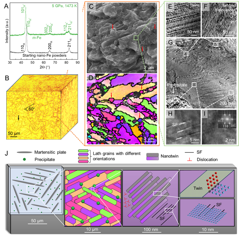
Figure 4. (A) XRD pattern of m-Fe synthesized at 5 GPa and 1,473 K. (B) 3D optical micrograph of a m-Fe cube with etched surfaces. (C-D) SEM and EBSD observations of a typically selected zone in (B). (E–I) Enlarged portions of a framed region in (C). (J) Schematic diagrams of hierarchically structured martensites in m-Fe.
High-pressure sintering at 5 GPa and 1473 K transformed the nano-Fe powders into single-phase martensitic Fe bulk samples, showing obvious XRD peak splitting (Figure 4A). These m-Fe bulk samples exhibited a hierarchically structured martensitic with different features across various scales (Figure 4B-4J).
At the microscale, the samples consist of martensitic plates and matrix. At smaller scales, the martensitic matrix is composed of laths and uniformly distributed spherical precipitates, while the nanometric laths contain high-density nanotwins, tangled dislocations, and bundles of SFs. At the atomic scale, abundant twin boundaries, SFs, and dislocations are abundant. All these spatial obstacles interact across scales to inhibit the dislocation slips and strengthen the material from both macroscopic and microscopic levels.
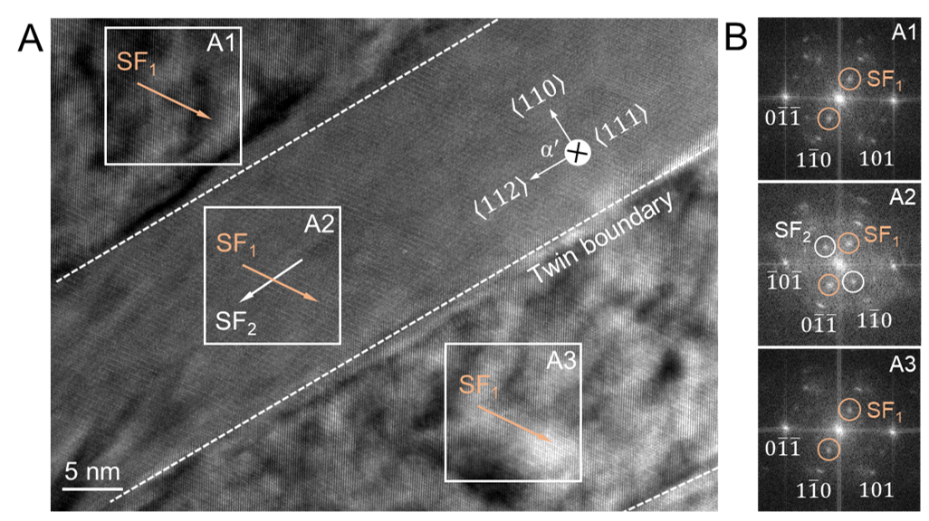
Figure 5. (A) TEM images of nanotwins and SFs in m-Fe. The yellow and white arrows represent two differently oriented SFs (i.e., SF1 and SF2 with a 60º angle). (B) FFT images of selected areas in (A) as denoted by A1, A2 and A3, respectively.
The two adjacent nanotwin domains of m-Fe exhibit different dislocation densities, with each domain containing different SFs identifiable through diffraction spots (Figure 5). One domain primarily features unidirectionally oriented SFs (i.e., SF1), while the other contains two differently oriented SFs (i.e., SF1-SF2). Such interesting twins are produced due to complex phase transitions in the associated domains under high-pressure and high-temperature conditions.

Figure 6. (A) Vickers hardness vs. load. for m-Fe and other references of HSS, T304, and SPS-Fe. (B) Plot of Vickers hardness of m-Fe and various reference steels vs. concentration of solute atoms.
The indentation experiments show that m-Fe has an unprecedentedly high load-invariant hardness of ~9.0 GPa, which is more than 20% and 190% greater than that of high-speed steels (HSS, ~7.5 GPa) and type-304 steels (T304, ~3.1 GPa), respectively (Figure 6). The apparent shrinking of indentation edges in m-Fe suggests its superior elastic recoverability over HSS.
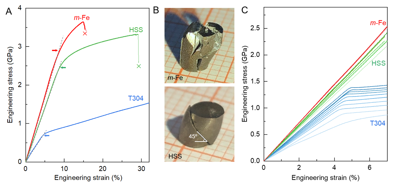
Figure 7. (A) Engineering stress vs. strain determined by uniaxial compression experiments. Arrows denote yield points. (B) Optical images of fractured samples. (C) Cyclic compression measurements with ten loading cycles.
Uniaxial compression experiments show that m-Fe achieves a yield strength of 2.9 GPa and an ultimate strength of 3.7 GPa, representing increases of 21% and 12% over HSS, respectively (Figure 7A). Unlike HSS, which fractures along a 45º-angle shear plane, m-Fe displays a unique zigzag fracture pattern due to nanoprecipitates that block shear deformation (Figure 7B). After ten cycles of compression at a maximum strain of 7% (Figure 7C), m-Fe shows a nearly invariant stress-strain relation, confirming its robust elastic recoverability. Remarkably, the yield strength of m-Fe is predominated by elastic stiffness, as further demonstrated by both the fracture-induced spark splashing and bouncing ball experiments (Videos 1-2).
Video 1. Bouncing ball experiments of micro-Fe, T304, HSS, and martensitic m-Fe.
Video 2. Compression experiments of m-Fe.

Figure 8. (A) In situ high-pressure and high-temperature XRD data. (B-G) Schematic of phase evolutions during cooling with four different stages as denoted by I, II, III, and IV, respectively, corresponding to four phases in (A).
The researchers used in situ high-pressure and high-temperature XRD measurements to study the structural evolution of m-Fe during cooling and decompression (Figure 8A). They found that the martensitic phase transition occurs in stages, beginning with partial dislocations initiating the formation of intermediate ε-Fe phase in the interior of each grain around 1373 K, which grows into large martensite plates with tens of micrometers (Figure 8B). As the temperature drops below 1373 K, smaller micrometric ε-Fe laths form at a slower growth rate due to increased pressure nonhydrostaticity (Figure 8C). At approximately 1173 K, new ε-Fe nuclei appear at lath boundaries, where plentiful crystalline imperfections encourage nucleation, forming nanometric bands as they grow (Figure 8D).
At around 973 K, the existing ε1-Fe bands intersect, inducing ε-to-α transitions that produce alternating ε-α layers, which are the precursors of nanotwins (Figure 8E). By 300 K, the ε-Fe becomes metastable and transforms into γ-Fe, while decompression transforms α-Fe into martensitic α’-Fe, and γ-Fe transitions through intermediate ε-Fe phases into α-Fe (Figure 8F-8G). This process explains the two distinct “texture” structures of the nanotwin domains, which arise from differing phase transitions and dislocation densities (Figure 5). The study also found that nanoprecipitates originate from the initial nano-Fe powders used in the experiments (Figure 3B).
The success in unveiling the detailed kinetics and dynamics of martensitic transition in Fe gives great insights into the underlying transition mechanisms, which would also provide important guidance for the design and production of ultrastrong steels and ceramics.
Chao Gu, a former postdoctoral fellow in Shanmin Wang’s group and currently an Assistant Research Fellow at the Quantum Science Center of Guangdong-Hong Kong-Macao Greater Bay Area, is the first author of the paper. Associate Professor Shanmin Wang is the corresponding author. Additional collaborators include Dr. Haiyan Chen from Stony Brook University and Professor Yusheng Zhao from Eastern Institute of Technology. The Department of Physics at SUSTech & the Quantum Science Center of Guangdong-Hongkong-Macao Greater Bay Area are the affiliated institutions of this work.
Paper link: https://doi.org/10.1073/pnas.2408119121
To read all stories about SUSTech science, subscribe to the monthly SUSTech Newsletter.
Proofread ByAdrian Cremin, Yingying XIA
Photo ByDepartment of Physics