Ptychography is a powerful lensless microscopy technique applicable to both X-rays and electrons, garnering widespread interest in materials science and biology. However, the high requirements for flux and coherence restrict ptychography primarily to large-scale facilities like synchrotrons. There has been a strong push to adapt this powerful technique for use with compact laboratory light sources, which face challenges due to their low temporal coherence (broadband) and flux of tabletop sources.
To date, most numerical methods for broadband reconstruction have assumed non-dispersive samples. In practice, however, many specimens exhibit wavelength-dependent behavior under broadband illumination, invalidating these models and preventing high-quality image recovery.

A research team led by Associate Professor Fucai Zhang from the Department of Electronic and Electrical Engineering at the Southern University of Science and Technology (SUSTech) has reported a novel ultra-broadband ptychography method tailored for dispersive specimens.
Their work, entitled “Ultra-broadband ptychography for dispersive samples,” has been published in Nature Communications.
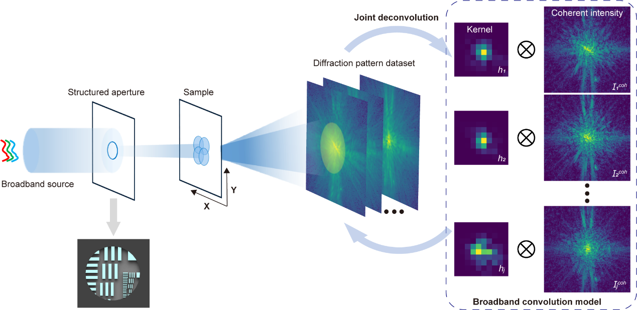
Figure 1. Schematic illustration of the ultra-broadband ptychography imaging principle
The researchers introduced an ultra-broadband ptychography method suitable for dispersive samples, addressing one of the core challenges not resolved in the current broadband robust algorithms. By combining structured illumination with a newly proposed joint deconvolution algorithm, the method enables high-resolution, high-fidelity quantitative phase recovery of dispersive samples without any monochromator-based filtering.
Using a light source with up to 60% bandwidth (400–1000 nm), the team successfully reconstructed high-quality, detailed images of a biological ant specimen, achieving a resolution error of only 7% relative to the theoretical limit. Further validation under a tabletop high-harmonic EUV source (46 nm) confirmed the method’s effectiveness for laboratory-scale broadband X-ray imaging.
This work paves the way for high-performance, laboratory-scale X-ray diffraction imaging and is poised to make a significant impact on chip inspection, materials characterization, and pharmaceutical development.
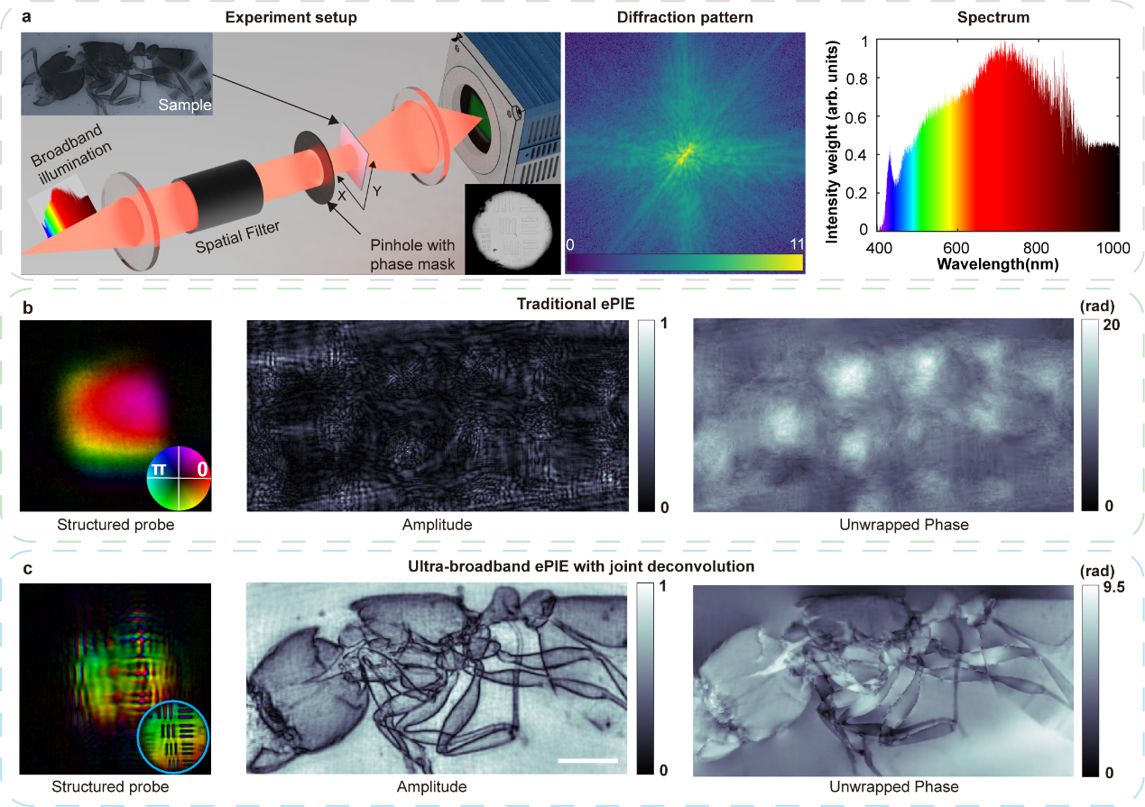
Figure 2. Imaging results of the ant sample under 60% bandwidth illumination
Ph.D. candidate Huixiang Lin is the first author of the paper, and Associate Professor Fucai Zhang is the corresponding author.
Paper link: https://www.nature.com/articles/s41467-025-61339-3
To read all stories about SUSTech science, subscribe to the monthly SUSTech Newsletter.
Proofread ByAdrian Cremin, Yifei REN
Photo ByDepartment of Electronic and Electrical Engineering


