On November 5, 2026, the research group of Professor Ning JIA, from the Department of Biochemistry and the Institute of Homeostatic Medicine, in the School of Medicine at the Southern University of Science and Technology (SUSTech), along with collaborators, published a study online in Molecular Cell titled “Bacterial 2’,3’-cGAMP signaling induces membrane splitting in antiphage CBASS immunity.” This work reveals a new bacterial antiphage immune mechanism, expands our understanding of the connection between bacterial and human innate immune signaling, and provides fresh ideas for developing novel immune-regulatory tools.
The arms race between bacteria and bacteriophages has persisted for billions of years. Through long-term co-evolution, bacteria have evolved a variety of sophisticated immune defense systems. Among them, the cyclic-oligonucleotide-based antiphage signaling systems (CBASS) are evolutionarily related to the mammalian cGAS-STING innate immune pathway: both rely on cGAS/DncV-like nucleotidyltransferases (CD-NTases) to synthesize cyclic oligonucleotide second messengers from nucleotide substrates, which are then converted into antiviral responses by downstream effector proteins. However, how bacteria sense phage invasion and activate CBASS systems, and how the resulting cyclic nucleotide signals drive effector proteins to mount antiviral defense, remain largely unexplored.
The research team revealed a new immune mechanism of the CdnB-Cap14 system in Bacillus thuringiensis and how phages evade it. CdnB senses phage DNA signals and produces 2’,3’-cGAMP, a signaling molecule identical to the one in the human innate immune cGAS-STING pathway. This activates the effector protein Cap14, which forms special fiber structures that split the cell membrane into two separate lipid bilayers, killing the infected bacteria and stopping the phage from spreading. Meanwhile, phages use the Acb2 protein to bind and sequester 2’,3’-cGAMP, evading the bacterial immune response.
The CdnB-Cap14 system is made up of the cyclic dinucleotide synthase CdnB and the effector protein Cap14. It’s a type I CBASS system and can produce the second messenger 2’,3’-cGAMP, just like the cGAS-STING pathway in mammals. CdnB is made by fusing an N-terminal CD-NTase domain with a C-terminal AGS-C domain, while Cap14 has an N-terminal transmembrane module and a C-terminal SAVED domain. However, there’s still a lot we don’t know about how CdnB gets activated to make 2’,3’-cGAMP and how this signal turns on Cap14 to defend against phages.
The research team first found that the CdnB-Cap14 system confers broad resistance against a diverse panel of bacteriophages (Figure 1A); infected bacteria sacrifice themselves to trap phages intracellularly and protect the surrounding population, a hallmark of “Abortive infection” (Abi). Deletion of CdnB or Cap14, or mutation of the catalytic DID motif to AIA, completely abolished resistance. In vitro, when purified CdnB was incubated with T2 phage genomic DNA together with ATP and GTP, thin-layer chromatography detected the 2’,3’-cGAMP product only when all three components were present; liquid chromatography and tandem mass spectrometry further confirmed its migration behavior and mass-to-charge ratio, and the reaction was strictly Mn2+-dependent. These results are corroborated by two recent studies published in Cell Host & Microbe and Cell (Figure 1B). The team solved the crystal structures of the N-terminal CD-NTase-domain truncation (CdnBΔC) in the apo state and in complex with ATP and GTP (Figure 1C). Based on structural analysis combined with in vitro biochemical assays, the team found that CdnBΔC itself possesses constitutive cyclase activity, indicating that the activity of the CD-NTase domain is autoinhibited by the C-terminal AGS-C domain (Figure 1D).
The cyclase activity of CdnB is regulated not only by phage DNA and its C-terminal AGS-C domain but also finely tuned by intracellular nucleotide levels: the enzyme efficiently synthesizes the second messenger only at low nucleotide concentrations, whereas further elevation leads to loss of activity (Figure 1E). This threshold is far below the physiological intracellular concentrations of ATP and GTP, echoing the nucleotide depletion that accompanies phage infection. This suggests that nucleotide depletion also constitutes a physiological signal that releases autoinhibition, enabling CdnB to avoid inappropriate activation in uninfected cells while rapidly producing 2’,3’-cGAMP when the cell is under severe threat.
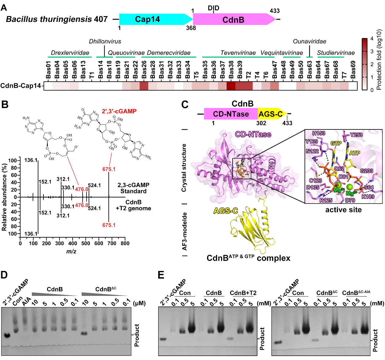
Figure 1. The CdnB-Cap14 system senses phage invasion signals and produces 2’,3’-cGAMP to defend against various phage infections. (A) The phage defense spectrum of the CdnB-Cap14 system; (B) CdnB can synthesize 2’,3’-cGAMP; (C) Crystal structure of CdnB bound to ATP and GTP; (D) CdnB without the AGS-C domain shows constitutive cyclase activity; (E) CdnB’s cyclase activity is regulated by phage DNA and nucleotide concentrations
The 2’,3’-cGAMP synthesized by CdnB then transmits the invasion signal to the downstream effector protein Cap14. Using cryo-EM single-particle analysis, the team solved the structure of the ligand-free Cap14 homodimer as well as the activated Cap14-2’,3’-cGAMP complex (Figure 2A). 2’,3’-cGAMP binds at the interface between the SAVED domains of adjacent protomers within the same strand and, acting as a “molecular glue,” drives Cap14 assembly from dimers into an open-ended, two-stranded filament with head-to-tail organization. Formation of the SAVED-domain filament is coupled to a dramatic rearrangement of the N-terminal transmembrane helices into a parallel, zipper-like, two-layered architecture. Cryo-EM density maps further showed that the continuous single layer of detergent micelle observed in the ligand-free state is split into two discrete micelles, leaving an evident density void between the two layers. This indicates that, in a physiological membrane environment, this conformational transition longitudinally partitions an originally continuous lipid bilayer into two independent bilayers.
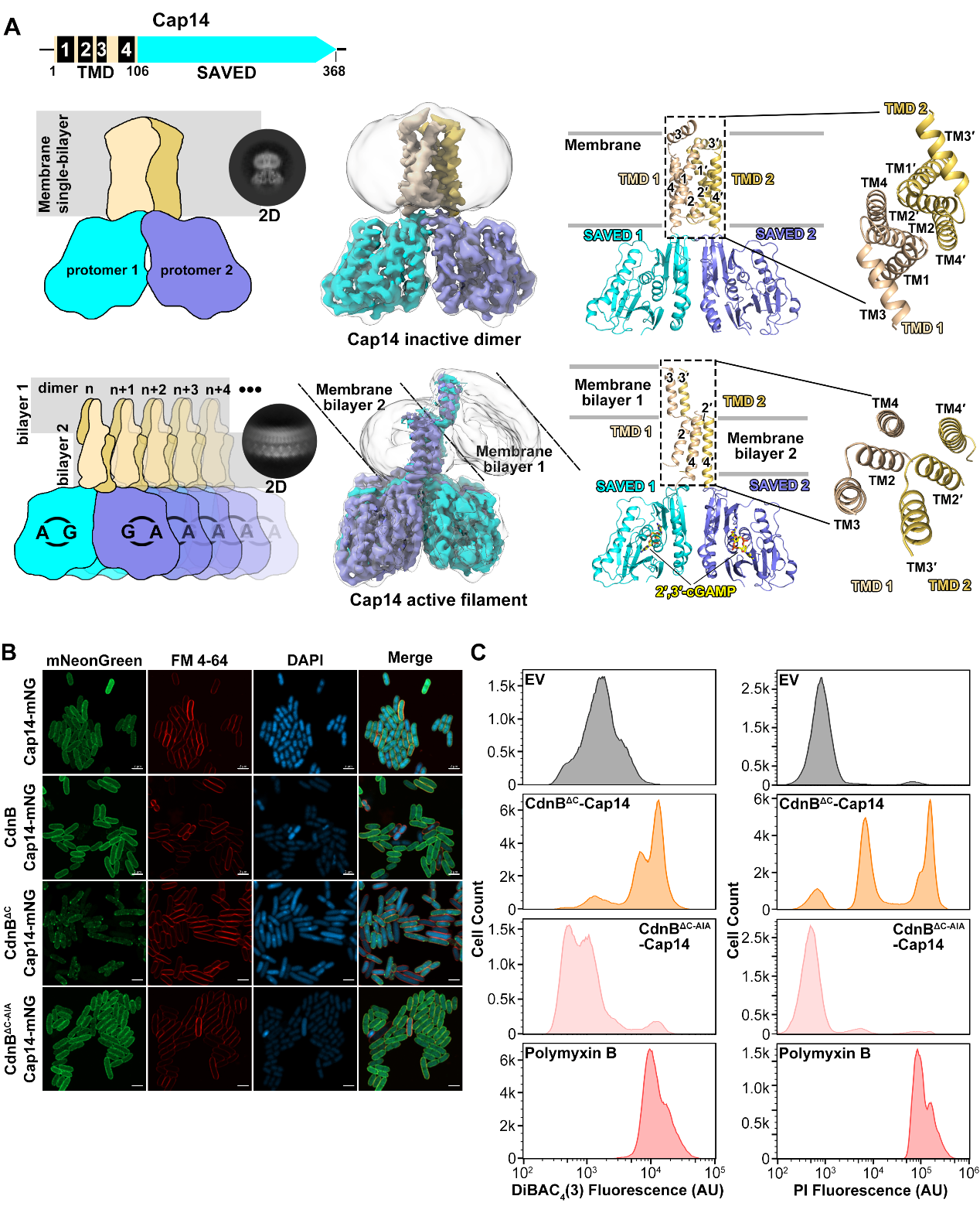
Figure 2. 2’,3’-cGAMP activates Cap14 to form zipper-like double-layered fiber structures and induces membrane splitting. (A) The structure of Cap14 dimers in the inactive state and the Cap14-2’,3’-cGAMP complex in the active state; (B) Activated Cap14 clusters on the cell membrane; (C) Activated Cap14 mediates cell membrane damage.
Further in vivo experiments showed that activated Cap14 forms prominent clusters on the cell membrane (Fig. 2B); mutations that disrupt the filament assembly interface or the hydrophilic–hydrophobic organization of the transmembrane region compromised resistance and cytotoxicity to varying degrees. Known CBASS effectors kill infected cells mostly by hydrolyzing membrane phospholipids, degrading nucleic acids, or depleting NAD+, whereas Cap14 neither forms pores nor hydrolyzes phospholipids; instead, it directly splits a continuous lipid bilayer into two discrete membranes, a previously undescribed mode of membrane damage. This mechanism is also supported by an independent study. The Gao group recently reported a closely related 3TM-SAVED effector in Bacillus thuringiensis and likewise observed 2’,3’-cGAMP-driven higher-order filament assembly accompanied by extensive rearrangement of the transmembrane region and membrane shearing (Cell 189.12 (2026): 3636–3650). Physiological assays further confirmed that activated Cap14 markedly compromises membrane potential and membrane integrity (Fig. 2C), showing that it induces cell death through this novel membrane-splitting mechanism and thereby defends against phage infection via abortive-infection self-sacrifice.
The study also revealed how phages escape CdnB-Cap14 defense. The Acb2 protein of T4 phage partially relieved the antiphage defense of the CdnB-Cap14 system (Figure 3B) and significantly alleviated the CdnBΔC-Cap14-mediated cytotoxicity when co-expressed with the constitutively active CdnBΔC and Cap14, whereas the Acb2 protein of T2 phage had no effect (Figure 3C). Replacing the key Tyr8 residue of T4-Acb2 with the corresponding Asp residue of T2-Acb2 completely abolished this inhibitory capacity. Biochemical assays further showed that, as the T4-Acb2 concentration increased, the free 2’,3’-cGAMP band progressively diminished; at an Acb2:cGAMP molar ratio above 5:1, the signal molecule was almost completely sequestered, with no cleavage products detected throughout, and the messenger could be released again upon heat denaturation (Figure 3D). These results demonstrate that T4-Acb2 is a catalytically inactive “sponge” protein that sequesters 2’,3’-cGAMP through direct binding, depleting the free messenger pool and blocking downstream signal transmission, thereby allowing the phage to escape.
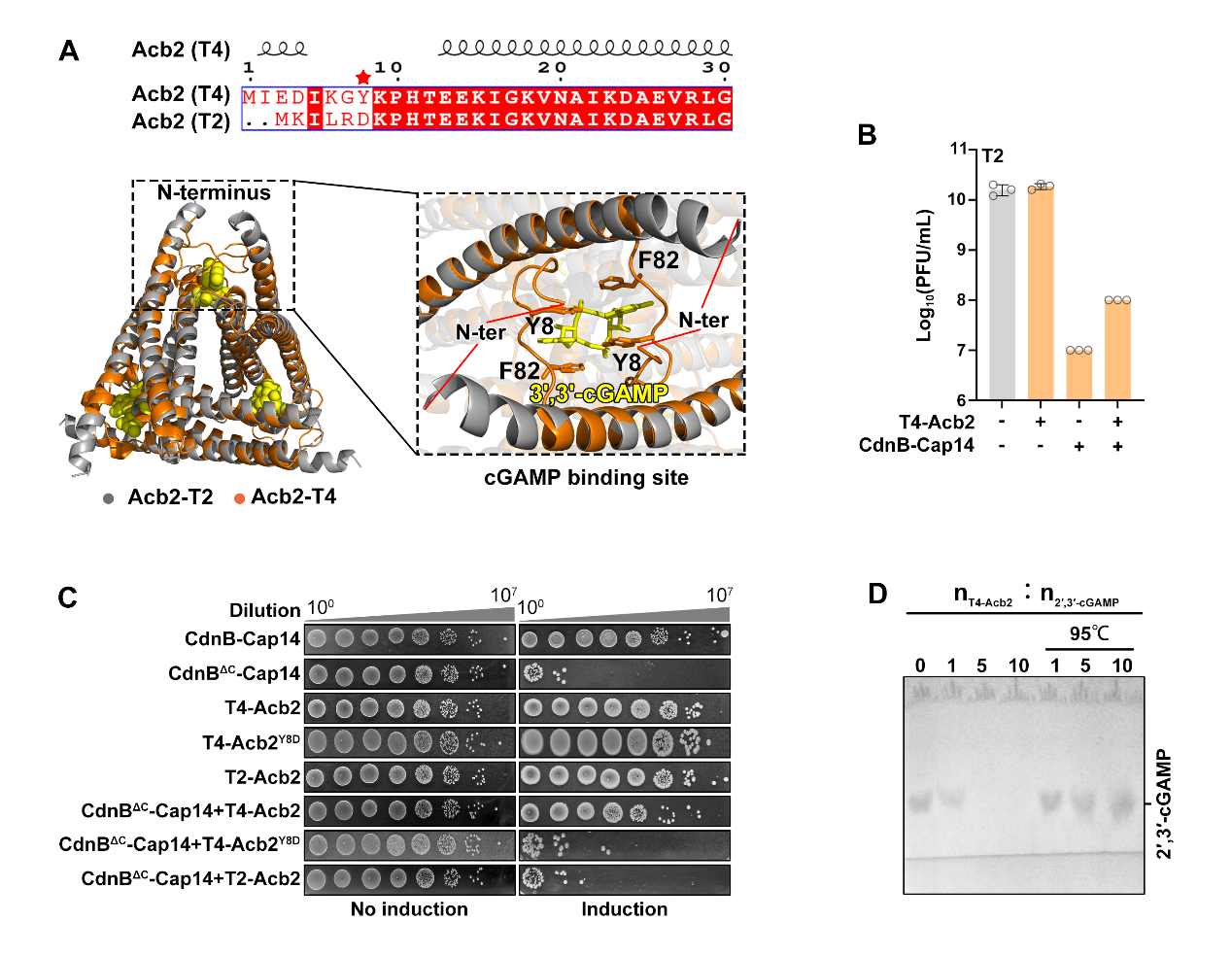
Figure 3. Acb2 protein helps phages escape the CdnB-Cap14 system by binding and sequestering 2’,3’-cGAMP. (A) Sequence and structure comparison of Acb2 proteins from T4 and T2; (B) T4-Acb2 protein helps phages escape the CdnB-Cap14 system; (C) T4-Acb2 suppresses the cytotoxicity mediated by activated Cap14; (D) T4-Acb2 protein binds and sequesters 2’,3’-cGAMP.
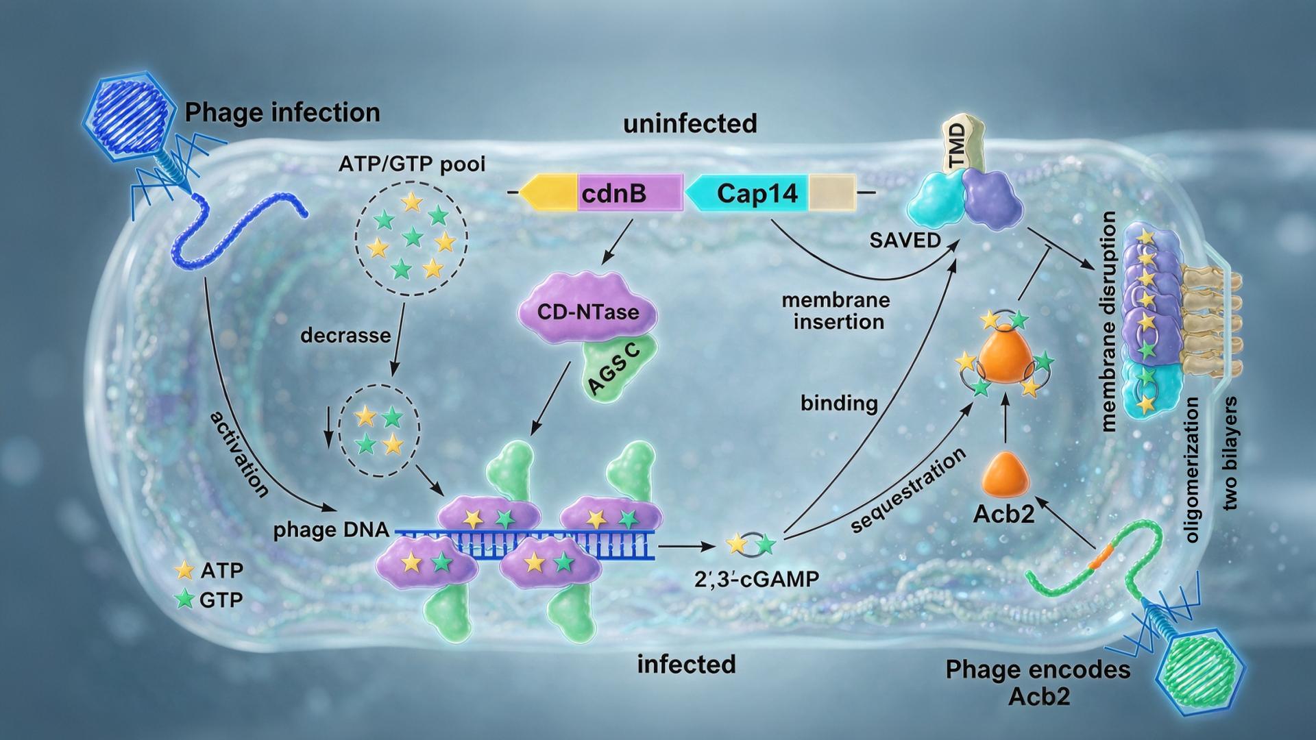
This study fully elucidates the molecular mechanism of the CdnB-Cap14 system, from CdnB autoinhibition, sensing of phage invasion signals, and 2’,3’-cGAMP synthesis, to Cap14 filamentous assembly and membrane splitting, and finally to phage immune evasion mediated by Acb2 (Figure 4). In uninfected cells, the cyclase activity of CdnB is locked in an autoinhibited state by its C-terminal AGS-C domain, and Cap14 is diffusely distributed on the cell membrane as a dimer. Upon phage invasion, phage genomic DNA together with infection-associated nucleotide depletion jointly release the autoinhibition, and CdnB synthesizes 2’,3’-cGAMP using ATP and GTP as substrates. This messenger binds the SAVED domain of Cap14 and drives its filamentous assembly, forcing the N-terminal transmembrane region to rearrange into a zipper-like two-layered architecture that splits the originally continuous lipid bilayer into two discrete membranes and disrupts membrane integrity; the infected bacterium consequently dies, and progeny phages are confined within the cell.
In the human cGAS-STING pathway, cGAS is likewise activated by DNA to synthesize 2’,3’-cGAMP; STING binds this messenger and undergoes higher-order oligomerization to recruit TBK1, initiating transcriptional immune activation. CdnB-Cap14 follows a highly similar “sense–synthesize–oligomerize” logic, yet culminates in a strikingly different effector outcome: not transcriptional reprogramming, but direct splitting of the membrane structure. These findings not only deepen our understanding of bacterial CBASS immunity but also strengthen the evolutionary connection between bacterial antiviral signaling and eukaryotic innate immunity. Of particular translational potential, the CdnBΔC truncation identified in this study is a compact, constitutively active module for 2’,3’-cGAMP synthesis whose product is chemically identical to the messenger produced by human cGAS; once engineered, it could enable sustained, controllable production of 2’,3’-cGAMP in mammalian cells, providing a molecular tool for modulating cGAS-STING pathway activity and offering key structural and mechanistic guidance for developing controllable intracellular 2’,3’-cGAMP production platforms, with potential applications in biotechnology and therapeutic engineering.
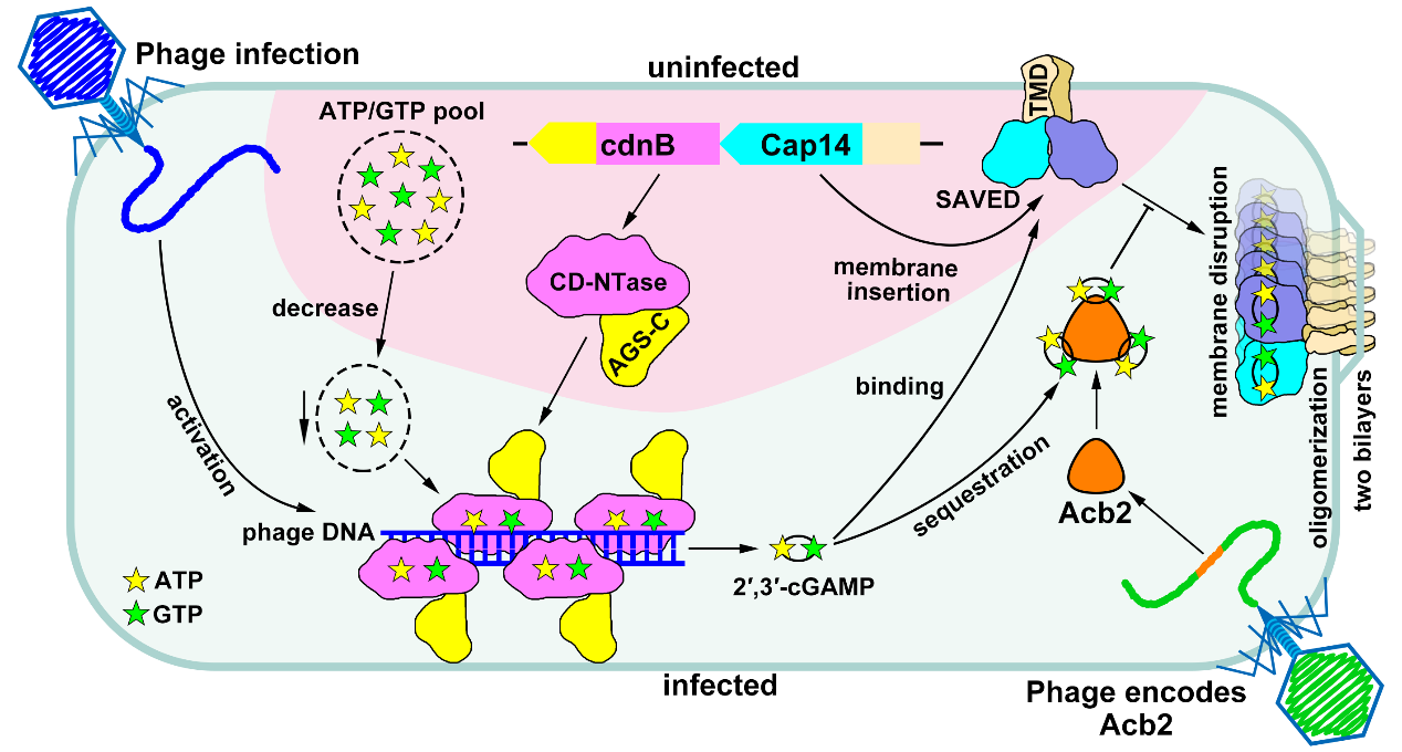
Figure 4. Mechanistic model of how the CdnB-Cap14 system senses phage invasion and induces membrane splitting to defend against phage infection.
PhD student Zhuolin LI from the School of Medicine at SUSTech and Wanbiao CHEN from the Shenzhen Institutes of Advanced Technology, Chinese Academy of Sciences, are co-first authors of the paper. Professor Ning JIA from the School of Medicine at SUSTech, Professor Chongyuan WANG from the Shenzhen Institutes of Advanced Technology, Chinese Academy of Sciences, and Research Assistant Professor Jun-Tao ZHANG from the School of Medicine at SUSTech are co-corresponding authors. SUSTech is the primary institution for the paper.
Paper Link: https://www.cell.com/molecular-cell/abstract/S1097-2765(26)00719-7
Proofread ByNoah Crockett, Junxi KE
Photo ByYan QIU