Getah virus (GETV) is a mosquito-borne arbovirus that belongs to the Semliki Forest group of the Alphavirus genus within the Togaviridae family. Since the first isolation from Culex gelidus mosquitoes in tropical rainforests of Malaysia in 1955, GETV’s infection spectrum has been expanding rapidly.
Professor Zheng Liu’s group from the Cryo-Electron Microscopy Center (Cryo-EM Center) at the Southern University of Science and Technology (SUSTech) collaborated with Professor Chuanqing Wang’s group in the College of Veterinary Medicine at the Henan Agricultural University (HAU) to publish an article on the structure of GETV.
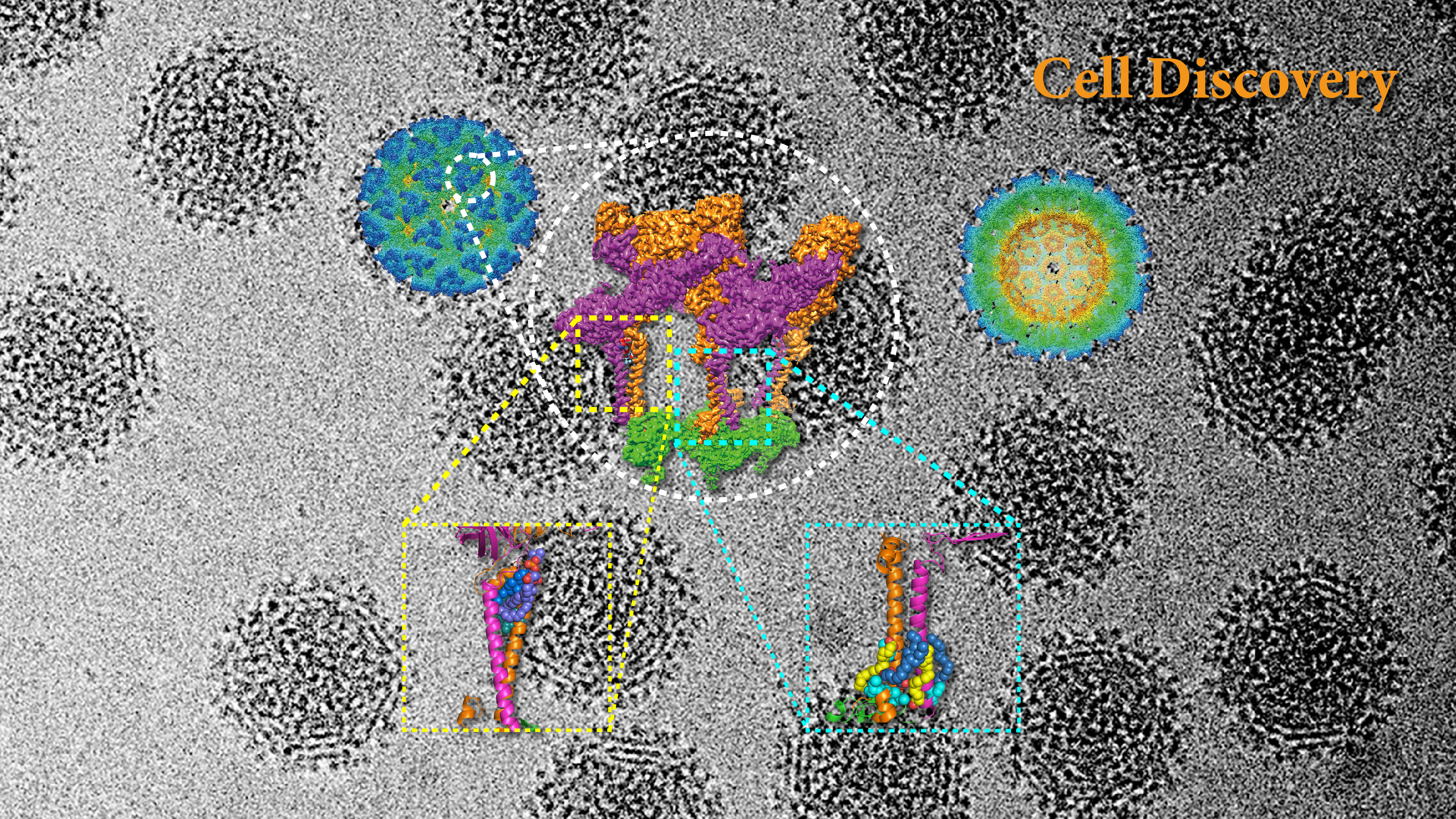
Their study, entitled “Structure of infective Getah virus at 2.8Å-resolution determined by cryo-electron microscopy”, was published in Cell Discovery, a high-quality journal covering all aspects of cell biology and molecular biology. In the study, the researchers utilized the newly constructed Biosafety Level 2 laboratory at Cryo-EM Center, to characterize the structure of mature GETV virions with infectious activity. The reported resolution of 2.8Å, is the highest resolution for an alphavirus to date.
The first outbreak of GETV among racehorses occurred in Japan in 1978, and outbreaks have re-emerged several times since then in Japan and India. GETV has been isolated from newborn piglets that died of acute disease and dead fetuses removed from infected sows in Japan.
The emergence of GETV in China was found in 2017 in swine farms, resulting in the death of the newborn piglets 5-10 days after birth and in pregnant sows exhibiting stillbirths or fetal mummies. Since then, infections have broken out in nearly ten provinces in China. In the past five years, the number of infected pigs in many farms has exponentially increased. Infected sows cause abortion, stillbirth, mummy stillbirth, and weak fetus. The infected 5-day-old piglets had a fever, diarrhea, and even death. GETV was also reported to infect beef cattle, blue foxes, and wild boars. Neutralizing antibodies of GETV have been detected in various vertebrate species, suggesting that domesticated animals can act as reservoir hosts.
Although the pathogenicity of GETV in humans has not yet been identified, seroepidemiologic investigations have shown that some individuals with a febrile illness have significantly higher GETV antibody titers than in healthy people, suggesting an association of GETV with human diseases. Considering lessons learned from the ongoing global pandemic of coronavirus disease (COVID-19) that resulted from the outbreak of severe acute respiratory syndrome coronavirus 2 (SARS-CoV-2), GETV presents a potential risk of becoming a zoonosis. Thus, plans and preparations should be made for the possibility of GETV transmission to humans.
Numerous glycosylation sites are identified on the viral envelope glycoproteins E1 and E2, revealing how they may participate in virus immune escape and host invasion. Several S-acylation sites are identified on the inner side of the transmembrane helical membrane for the first time, which greatly stabilized the transmembrane assembly of E1 and E2. Cholesterol and phospholipid molecules were identified in the hydrophobic pocket outside the transmembrane helical membrane for the first time, which played a synergistic role in stabilizing the hydrophobic pocket in the virus envelope.
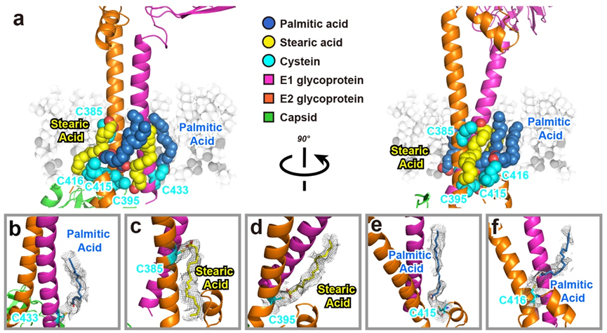
Figure 1. S-acylation were firstly identified on the inner side of the transmembrane helical membrane
Based on the high-resolution structure of the study, the research team applied the latest artificial intelligence technology to screen the zinc library, which contains more than seven million compounds. Nine compounds that potentially bind to the virus have been selected. At present, the cell viability assay and anti-virus experiments at cell and animal levels are in progress. As GETV widely infects livestock and causes disease, developing drugs against GETV significantly impacts China, the world’s largest livestock raising country.
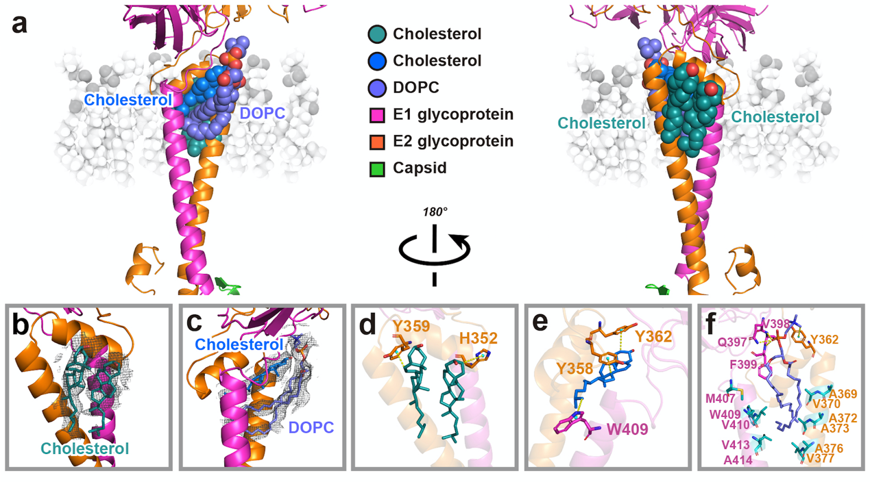
Figure 2. Cholesterol and phospholipid molecules were identified for the first time in the hydrophobic pocket outside the transmembrane helical membrane
Research Assistant Aojie Wang, Dr. Congcong Liu of the Second Affiliated Hospital of SUSTech (Shenzhen Third People’s Hospital), and Dr. Feng Zhou of HAU are the co-first authors of this paper. Prof. Zheng Liu from the Cryo-EM Center at SUSTech and Prof. Chuanqing Wang from HAU are the co-corresponding authors. SUSTech is the leading affiliate of this paper.
Dr. Ruxi Qi, Dr. Sheng Liu, Dr. Yuanzhu Gao, and Mr. Lutang Fu from the Cryo-EM Center also assisted in this research. The Cryo-EM data collection and image processing were completed in the Cryo-EM Center. The work was supported by the National Natural Science Foundation of China (NSFC) and the Startup Fund from SUSTech.
Paper link: https://www.nature.com/articles/s41421-022-00374-6
To read all stories about SUSTech science, subscribe to the monthly SUSTech Newsletter.
Proofread ByAdrian Cremin, Yingying XIA
Photo By