As the “powerhouses” of the cell, mitochondria are vital to cellular function and disease development. However, traditional mitochondrial biopsy techniques rely on fluorescent labeling, a method associated with photodamage, cytotoxicity, and interference with downstream analyses. Furthermore, existing intracellular manipulation techniques continue to face numerous challenges in terms of precision, minimal invasiveness, and operational efficiency.
Associate Professor Chengzhi Hu’s research team from the Department of Mechanical and Energy Engineering at the Southern University of Science and Technology (SUSTech), in collaboration with Assistant Professor Hongri Gu at the Hong Kong University of Science and Technology (HKUST), has made new progress in the field of robot-assisted, label-free intracellular mitochondrial biopsy.
Their related findings have been published in Science Advances under the title “Label-free robotic mitochondrial biopsy.”

Figure 1. Nanoprobe structure, fabrication, and functions
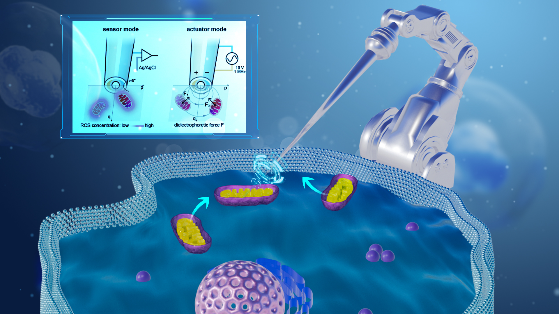
The researchers developed an automated, multifunctional nanoprobing platform capable of label-free extraction of mitochondria from living cells with high spatiotemporal resolution. The nanoprobe integrates two individually addressable nanoelectrodes that perform electrochemical detection of reactive oxygen and nitrogen species (ROS/RNS) produced by mitochondrial metabolism, followed by dielectrophoretic trapping, manipulation, and extraction of mitochondria.
The platform demonstrates several key technological advantages. It features multifunctional integration, as the same nanoprobe performs dual functionality in both sensing and actuation, enabling it to detect mitochondria as well as capture and extract them. It’s label-free detection approach eliminates the need for fluorescent labeling, thereby avoiding photodamage and cytotoxicity and facilitating a wide range of subsequent biological analyses.
The system also provides high spatiotemporal resolution, achieving a temporal resolution of 1 kHz and a spatial resolution of 1 μm when functioning as an sensor, and a spatial resolution of 1.8 μm when acting as an actuator. Moreover, its low invasiveness is made possible by a miniaturized tip and precise robotic control that allows for highly minimally invasive cell manipulation, with a demonstrated cell survival rate as high as 96%.
The research team further utilized this technology to achieve intercellular mitochondrial transplantation, observing that the transplanted mitochondria successfully integrated into the mitochondrial network of the recipient cells. This achievement not only provides a powerful tool for investigating mitochondrial function and related disease mechanisms but also opens up new technological pathways for single-cell surgery and cell therapy.

Figure 2. Automated robotic detection and manipulation of mitochondria

Figure 3. Robotic mitochondrial transplantation
Ph.D. candidate Yanmei Ma from the Department of Mechanical and Energy Engineering at SUSTech and joint Ph.D. candidate Weikang Hu of SUSTech and the City University of Hong Kong are co-first authors of the paper. Associate Professor Chengzhi Hu and Assistant Professor Hongri Gu are the corresponding authors, with SUSTech serving as the primary affiliated institution.
Paper link: https://www.science.org/doi/10.1126/sciadv.adx4289
To read all stories about SUSTech science, subscribe to the monthly SUSTech Newsletter.
Proofread ByAdrian Cremin, Yuwen ZENG
Photo ByDepartment of Mechanical and Energy Engineering


