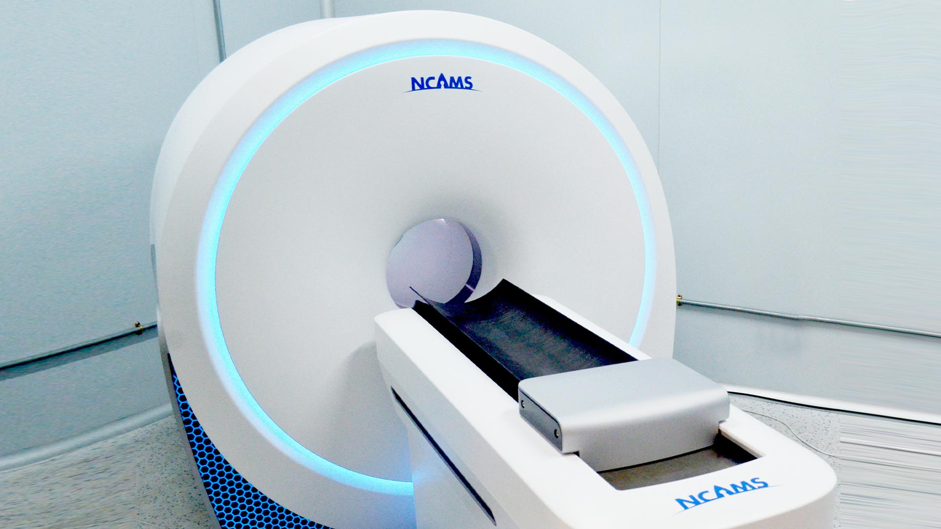
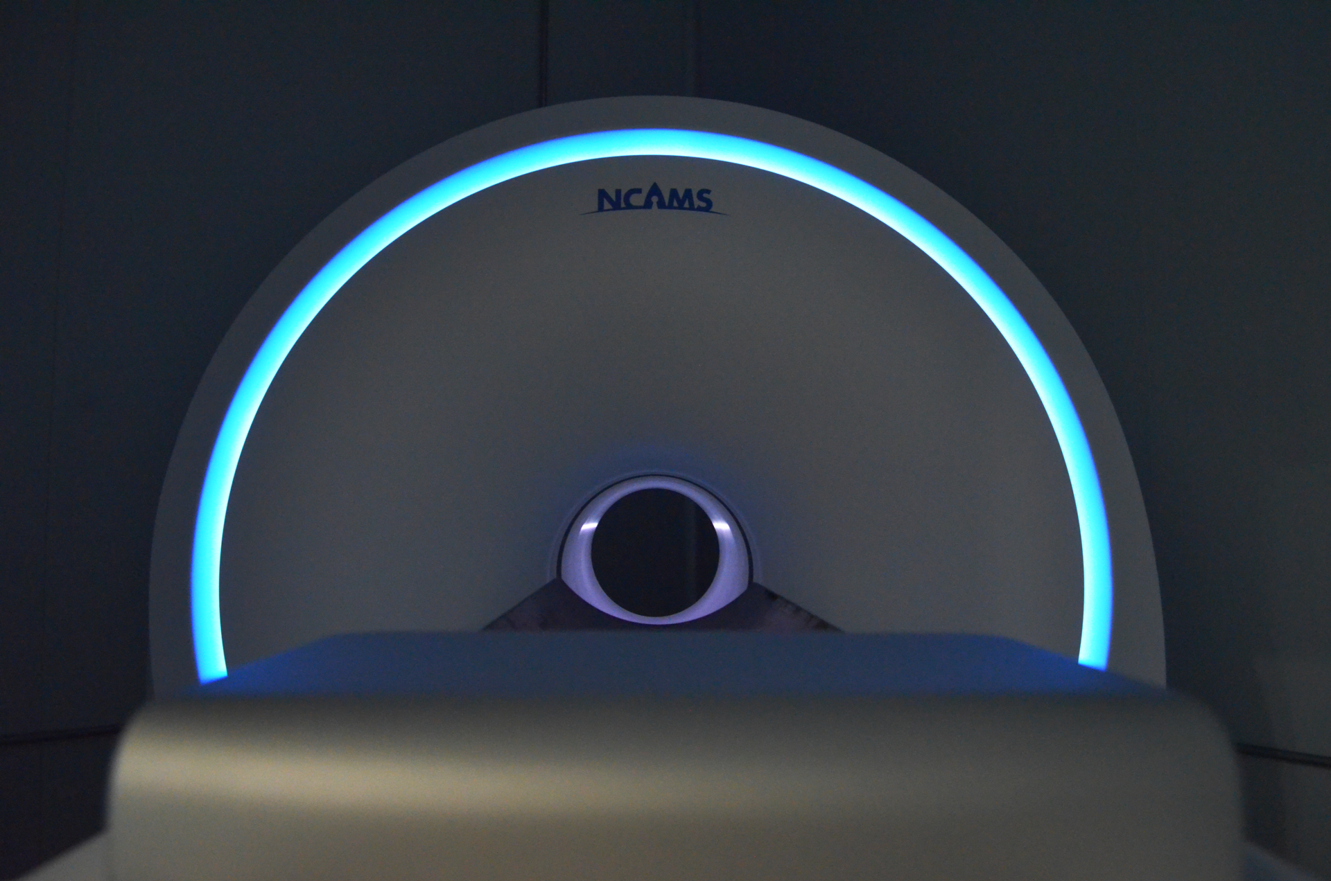
The CT Imaging Engineering Laboratory of the National Center for Applied Mathematics Shenzhen (NCAMS) has successfully developed the first orthopedic spectral cone-beam CT prototype.
The team of researchers from the Southern University of Science and Technology (SUSTech), led by Fuquan FANG, Chair Professor of the Department of Mathematics and Academician of the Chinese Academy of Sciences (CAS), has made an important breakthrough in high-end spectral CT algorithm and technological innovation, and has filled the gap in the area of developing orthopedic spectral quantitative cone-beam CT imaging equipment.


Team members Professor Xing ZHAO and Professor Yunsong ZHAO noted that the device is based on advanced photon counting multi-spectral CT imaging technology, with a spatial resolution better than 50 μm, which can achieve low-dose, high-definition images of fine structures in cortical and cancellous bones.
The imaging can reveal mechanical and functional changes in orthopedic diseases through the analysis of bone trabeculae.
At the same time, multi-tissue recognition is made possible by the imaging process modeling and high-precision solution algorithms, a new solution with fully independent intellectual property rights. This makes it easier to tell the difference between muscle, intramuscular fat, cartilage, bone, and other tissue structures, for more accurate diagnosis.
Currently, the team has used the device to carry out scientific research on preclinical animal models with bone diseases, which will then be used in researching and diagnosing human peripheral bone and joint diseases.
In the future, NCAMS will continue to focus on advancing precision medical applications through computing and modeling, promoting in-depth cooperation between academia, industry, and research, and supporting mathematical talents to carry out innovative and ground-breaking scientific and technological research.
Proofread ByYingying XIA
Photo By