On July 13, 2026, the research team led by Associate Professor Yan ZHAO at the School of Life Sciences, Southern University of Science and Technology (SUSTech), published a mechanistic study in the Journal of Cell Biology titled “PISD acts as a switch between lipophagy and fatty acid transfer to mitochondria.”
Lipid droplets (LDs) are essential intracellular organelles that store energy. Under stress conditions such as starvation, neutral lipids stored in LDs are broken down into fatty acids (FAs) and transported to mitochondria for β-oxidation to produce energy. At the same time, cells also activate lipophagy, a process in which autophagosomes engulf lipid droplets and deliver them to lysosomes for degradation. How these two pathways are coordinated to ensure efficient lipid utilization and maintain energy homeostasis has remained unclear.
The study reveals a specific isoform of phosphatidylserine decarboxylase (PISD) localized on lipid droplets (LDs), termed PISD-LD, that serves as a critical molecular switch coordinating lipophagy and fatty acid (FA) transport. Under starvation conditions, PISD-LD mediates the physical interaction between lipid droplets and mitochondria, facilitating FA transfer and subsequent β-oxidation for energy production. Concurrently, PISD-LD suppresses lipophagy, ensuring that lipids are preferentially channeled to mitochondria for energy generation. This regulatory mechanism maintains the fine-tuned balance between efficient energy production and lipid homeostasis under nutrient-deprived conditions.
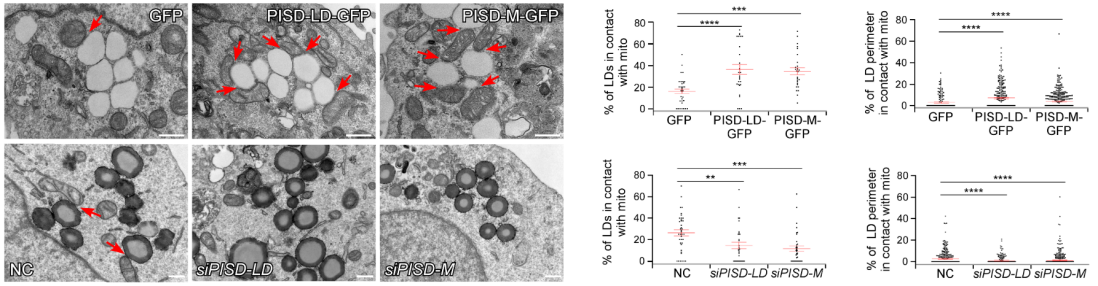
Figure 1. TEM imaging showed that LD–mitochondria contacts were markedly enhanced upon overexpression of PISD-LD-GFP or PISD-M-GFP, whereas knockdown of either isoform substantially diminished these contacts.
PISD-LD and the mitochondria-localized isoform PISD-M are generated from the same gene via alternative splicing. They possess distinct N-terminal sequences that direct their respective localization to the lipid droplet surface and mitochondria. The research team found that PISDs mediate membrane contacts between lipid droplets and mitochondria (Figure 1). Specifically, PISD-LD and PISD-M interact with each other through specific amino acid sites, functioning as tethering proteins to facilitate the formation of LD-mitochondria membrane contact sites. At these contact sites, PISD further recruits the lipid transfer proteins ATG2A and ATG2B, which utilize their lipid transfer domains to efficiently channel fatty acids from lipid droplets to mitochondria for β-oxidation. Meanwhile, PISD-LD negatively regulates lipophagy by binding to the lipophagy receptor Spartin, thereby competitively inhibiting the interaction between Spartin and the autophagy protein LC3. Upon prolonged starvation or impaired mitochondrial β-oxidation, PISD-LD undergoes proteasomal degradation, which relieves the inhibition on Spartin. Freed from this suppression, Spartin subsequently binds LC3, triggering the encapsulation of lipid droplets by autophagosomes and their delivery to lysosomes for degradation.
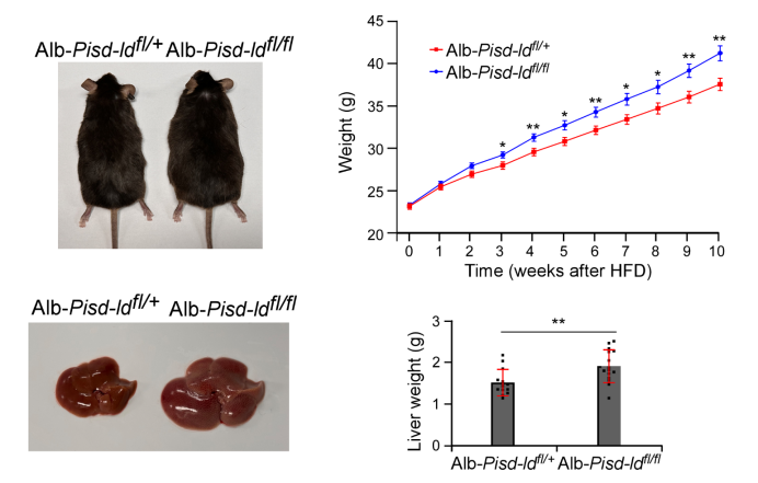
Figure 2. Liver-specific knockout of Pisd-ld in mice resulted in significantly increased body weight and liver weight.
To validate the physiological significance of PISD-LD, the research team generated a liver-specific Pisd-ld knockout mouse model. Under high-fat diet feeding conditions, the knockout mice exhibited significantly increased body weight and liver weight compared with control littermates (Figure 2). Serum and hepatic triglyceride levels were elevated, and liver function markers (AST/ALT) were markedly increased, indicating aggravated liver injury. Histological staining revealed substantial accumulation and enlargement of lipid droplets in the livers of knockout mice. Transmission electron microscopy analysis further demonstrated that Pisd-ld deletion led to a pronounced reduction in LD-mitochondria contacts. Collectively, these findings establish a critical role for PISD-LD in regulating LD-mitochondria contacts and lipid metabolism in the mammalian liver, offering novel insights into the understanding and treatment of metabolic diseases such as fatty liver disease.
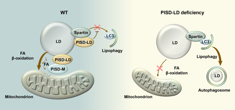
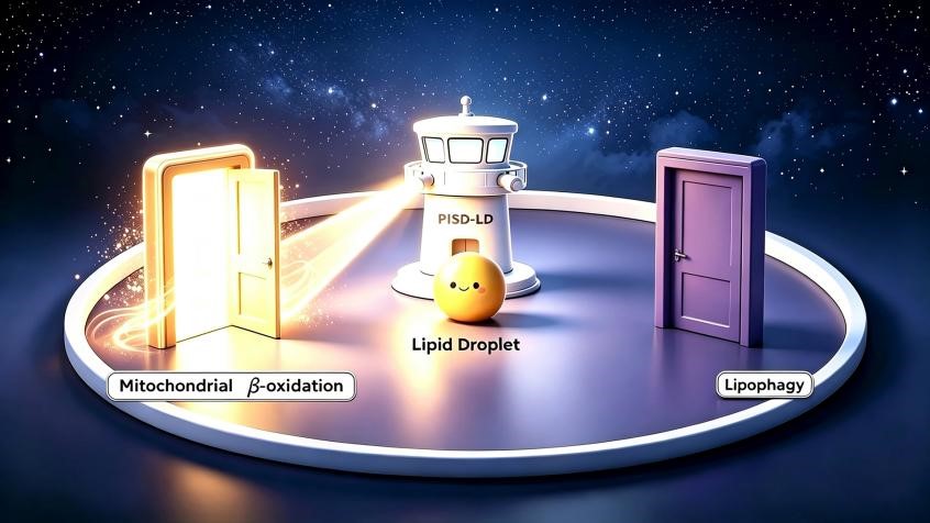
Figure 3. Schematic diagram illustrating the PISD-LD-mediated switch between fatty acid transfer and lipophagy.
PISD-LD exerts a dual role in coordinating lipid utilization. On one hand, it synergizes with PISD-M to mediate LD-mitochondria interactions and recruits the lipid transfer proteins ATG2A and ATG2B, thereby facilitating the transfer of fatty acids from lipid droplets to mitochondria for β-oxidation. On the other hand, PISD-LD binds to the lipophagy receptor Spartin, blocking its interaction with the autophagy protein LC3 and thereby suppressing lipophagy. Upon PISD-LD deficiency, Spartin is released and subsequently engages LC3 to initiate lipophagy, leading to lysosomal degradation of lipid droplets (Figure 3). This study not only elucidates a novel mechanism governing LD-mitochondria communication and lipophagic regulation but also provides important clues for understanding the pathogenesis of related metabolic disorders.
Associate Professor Yan ZHAO from the School of Life Sciences at SUSTech is the corresponding author of this paper, with SUSTech as the primary affiliation. Dr. Hanbing XUE (Research Assistant Professor in ZHAO’s lab), Ms. Xingwen HU (a recent master’s graduate from ZHAO’s lab), and Ms. Zhenni YANG (a doctoral student from Yunnan University) are co-first authors.
Proofread ByNoah Crockett, Junxi KE
Photo BySchool of Life Sciences