Assistant Professor Shujun CAI’s research team from the School of Life Sciences at the Southern University of Science and Technology (SUSTech), together with Associate Professor Ruoxi WANG’s team, published a study titled “In situ architecture of developmentally programmed mitophagy reveals ER-phagophore membrane continuity” in the international academic journal Autophagy. Using advanced imaging techniques like in situ cryo-electron tomography (cryo-ET), they analyzed the 3D structure of mitochondrial autophagy during fruit fly development and for the first time obtained in situ evidence showing direct membrane continuity between the endoplasmic reticulum (ER) and the phagophore membrane (also known as the isolation membrane). This provides new clues for understanding where the phagophore membrane comes from when large mitochondria are quickly wrapped up.
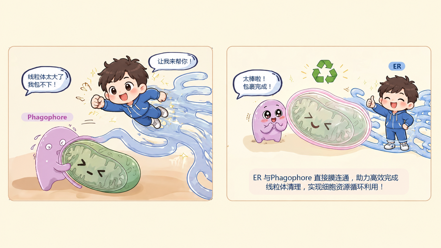
Autophagy is a highly conserved degradation process in eukaryotic cells. During autophagy, the cup-shaped phagophore gradually expands and engulfs the material to be degraded, then closes to form an autophagosome with a double-membrane structure, and finally fuses with the lysosome so that its contents are degraded. Mitophagy is the selective clearance of damaged or excess mitochondria, which is important for mitochondrial quality control, organism development, and cell homeostasis. During mitophagy, the phagophore membrane needs to rapidly extend along the surface of large mitochondria within minutes, which requires a continuous and abundant supply of membrane lipids.
There are currently two main models regarding the origin of phagophore membranes: one suggests that membranous vesicles can supply membrane materials to the phagophore membrane through transport and fusion; the other proposes that the endoplasmic reticulum can deliver lipids to the phagophore membrane at membrane contact sites via bridge-like lipid transfer proteins such as ATG2. It’s estimated that forming an autophagosome about 400 nanometers in diameter in roughly 10 minutes requires integrating around 3 million lipid molecules. However, the lipid transfer rate of ATG2 measured in vitro and its limited cellular abundance can’t fully explain the high flux of lipids needed for the rapid expansion of the phagophore membrane. Whether the ER can also supply membrane materials to the phagophore membrane through direct membrane continuity still lacks high-resolution in situ structural evidence under natural conditions.
To tackle this issue, the research team used mitochondrial autophagy (mitophagy) driven by developmental programs in the gut epithelial cells of pupal fruit flies as their study system, proposing a model in which the ER can form direct membrane-continuous structures with phagophore membranes. To observe the ultrastructure of cells in thick tissues as close to their natural state as possible, the team combined several cutting-edge techniques like Waffle high-pressure freezing, cryo-fluorescence localization, SOLIST, Cryo-FIB thinning, and Cryo-ET in order to achieve in situ 3D imaging of mitochondrial autophagy in the gut during fruit fly development.
Cryo-ET analysis showed that in some mitochondria undergoing the later stages of expansion, researchers observed small membrane openings about 600 to 800 square nanometers in size in the mitophagophore membranes. These openings are directly connected to the ER membrane network through narrow membrane necks, forming a truly continuous membrane structure rather than membrane contacts (Fig. 1). This finding provides the first in situ structural evidence of the ER being directly connected to phagophore membranes.
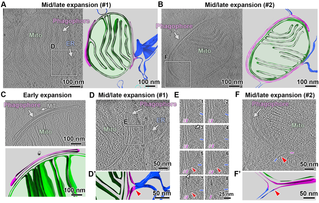
Fig. 1 Cryo-ET reveals that expanding mitophagophores form direct membrane continuities with the ER network.
To broaden the sampling range and further analyze the relationship between this structure and membrane expansion, the research team used room-temperature focused ion beam scanning electron microscopy (RT-FIB-SEM) for large-volume 3D imaging. In the control samples, 5 out of 49 mitophagophore membranes showed structures directly continuous with the endoplasmic reticulum. To further verify the link between this structure and membrane expansion, the team analyzed mutants of the key mitophagy regulator VPS13D that lack the UBA domain. In these mutants, many of the stalled, expanded mitophagophore membranes remained continuously connected to the ER (Fig. 2), indicating that this continuous membrane structure is dynamically regulated and closely tied to phagophore membrane expansion.
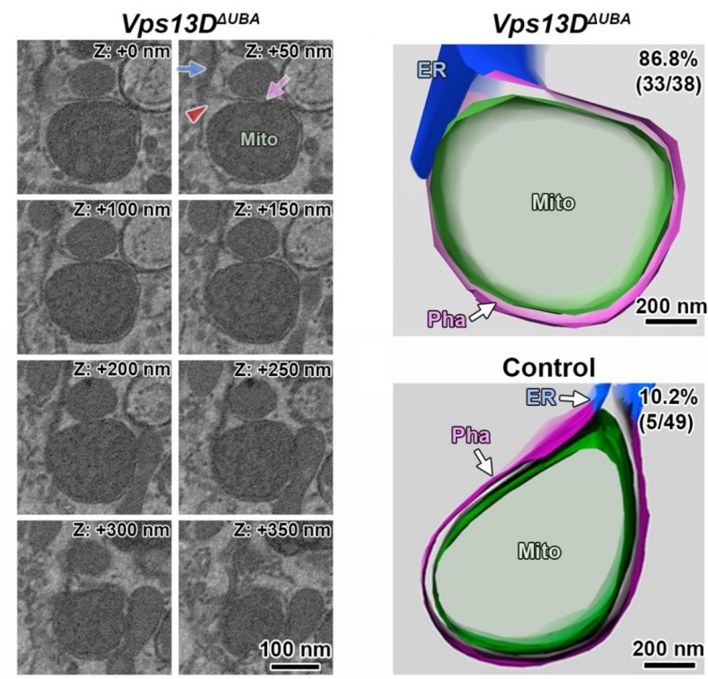
Fig. 2 In Vps13DΔUBA flies, a large number of mitophagophore membranes maintain membrane continuity with the ER.
This study, for the first time, obtained 3D structural evidence of the direct membrane continuity between the ER and phagophore membranes in a natural cellular environment. It proposes a new mechanism where the ER can directly supply membrane material to the mitophagophore membranes by establishing continuous membrane structures (Fig. 3). This finding enriches our understanding of the sources of autophagic membranes and provides a new framework for studying autophagosome formation. Whether this mechanism applies to other types or systems of autophagy, and how it coordinates with lipid transfer at membrane contact sites, still needs further investigation.
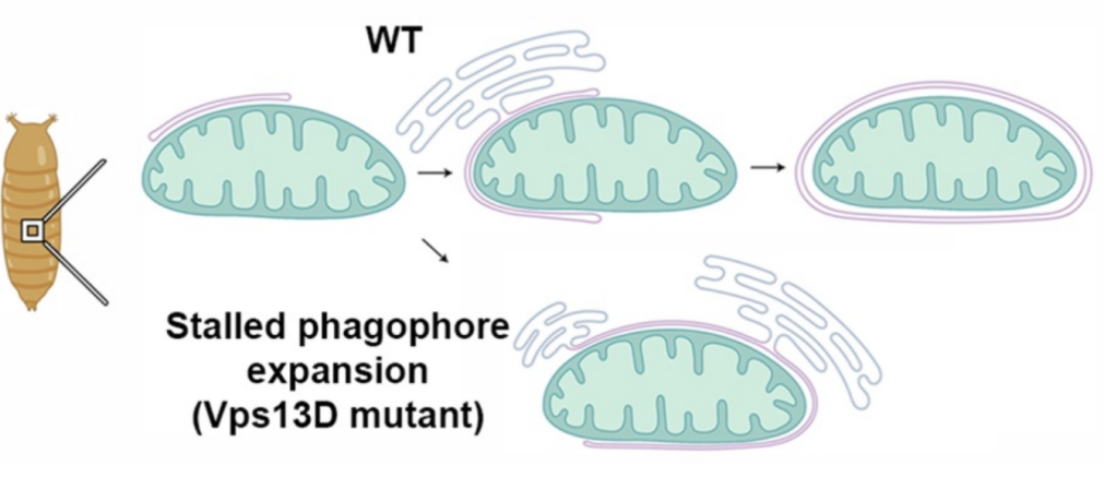
Fig. 3 A model of how mitochondrial autophagosomes form in the fruit fly gut during development.
Yufei HUANG, a Master’s student, and Tianyu ZHENG, a PhD student at the School of Life Sciences at SUSTech, are both co-first authors. PhD student Xiaofeng SUN also participated in the research and is listed as a paper author. Shujun CAI and Ruoxi WANG are co-corresponding authors, and SUSTech is the first affiliation of the paper.
Paper Link: https://doi.org/10.1080/15548627.2026.2657543
Proofread ByNoah Crockett, Junxi KE
Photo By